Eye Discharge in Dogs – What Does it Mean if Your Dog Has Runny Eyes?
Many dog owners will be familiar with occasional runny eyes in dogs. This is usually nothing to worry about. However, sometimes, runny eyes can be associated with more serious health conditions. Holidays4Dogs finds out more about eye discharge in dogs, what to look out for and when to be concerned.
There are several causes of watery eyes in dogs and, although common and generally not harmful, watery eyes can be a sign of a more serious disease.
Some of these acute conditions include;-
- Glaucoma – this is a chronic eye condition which leads to seriously limited vision, or blindness. Glaucoma refers to an increase in pressure within the inner eye. Symptoms often begin with itchy, watery eyes.
- Conjunctivitis – this can be a difficult condition to treat. Bacteria, or a virus can cause conjunctivitis. It is a condition that can cause considerable irritation to the eye. A dry atmosphere, or hairs around the eye can cause acute inflammation and discomfort.
- ‘Cherry Eye’ is a prolapsed gland of the third eyelid, which is located at the corner of the lower lid. It can be spotted easily as a red-coloured lump in the corner of the eye. This can cause complications if not treated, (usually surgery).
All these conditions are treatable. Therefore, if you notice eye discharge in your dog, it is important to take your dog to the vet.
Other possible causes might be;-
- Foreign body in the eye. If your dog’s eyes suddenly start weeping or watering, check for debris lodged within the lid, or the eye itself. Grass seeds are a common culprit for eye infections in dogs.
- Allergies are a potential cause of watery eyes – although dogs tend to have skin issues with allergies, it is possible the eyes too may be affected.
- Dry air, draughts, or windy conditions can also cause watery eyes, although this is often temporary.
Certain breeds of dogs can be more susceptible to eye conditions. Some of these breeds include short-nosed dogs such as bulldogs and toy breeds like Chihuahuas. In addition, large breeds, where the lower lid drapes down can also be susceptible, such as Bloodhounds.
Tears
Tears in the eye, play an important part in helping to maintain clear, healthy eyes and vision in animals. 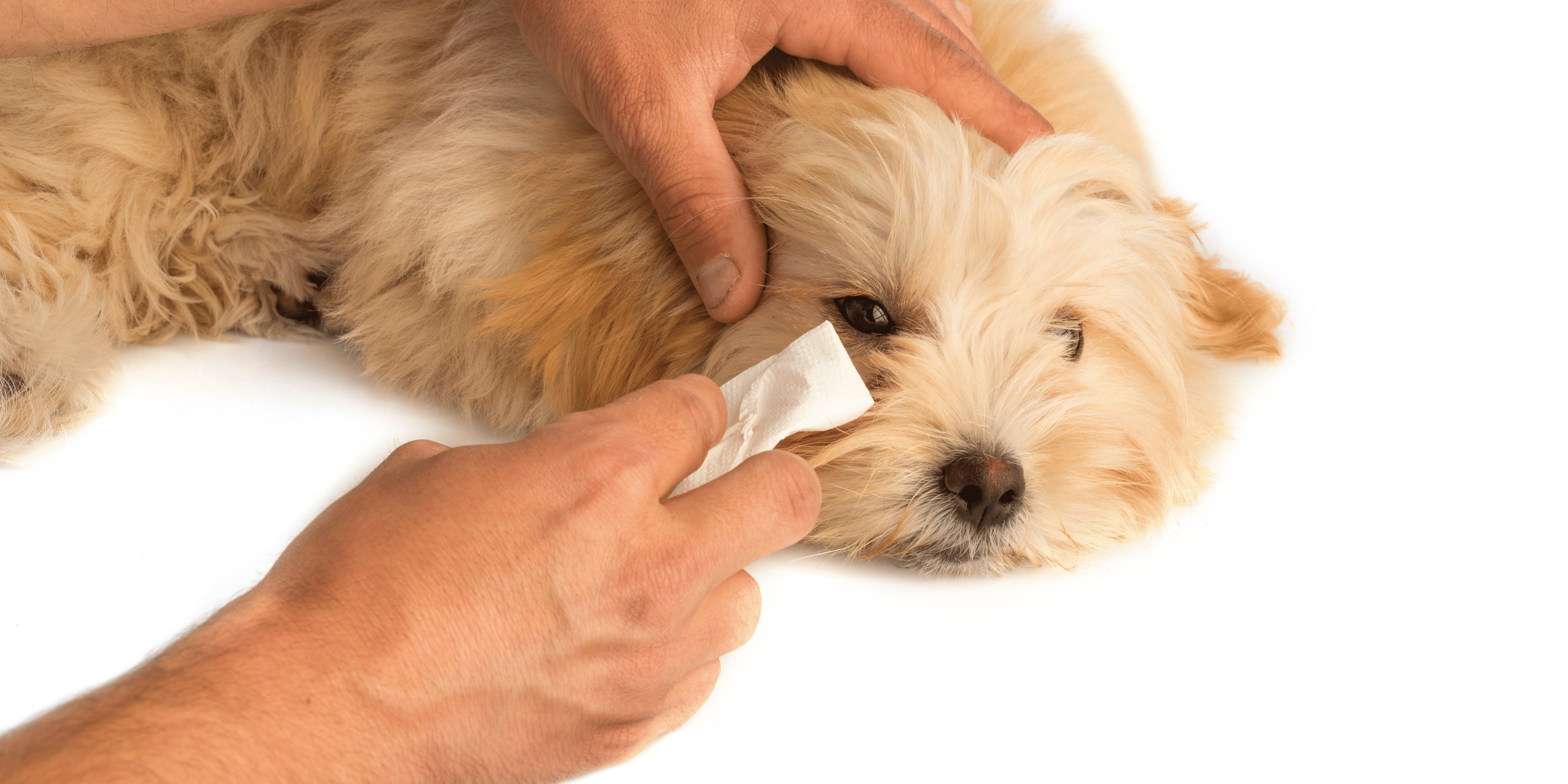 Not only do tears remove debris from the surface of the eye, but they also provide oxygen to the cornea, (the layer of clear tissue that covers the surface of the eye).
Sometimes the tubes, (or ducts) that tears drain through, can become temporarily blocked with dry tears, dust, dead cells and mucous. This can produce a crusty deposit around the eyes. In humans, many people refer to this as having ‘sleep’ in their eyes, as it often occurs after a long night’s sleep.
If your dog’s eyes are watering a lot this is usually the body’s way of trying to clear an obstruction – pollen or dust for instance. Watery eyes may also be a result of exposure to windy conditions. Isolated eye-watering, or eye discharge, generally clears up within 24 – 48 hours.
Discharge colour
If your dog has reddish-brown tear stains on his fur, (more noticeable in light-coloured dogs) this is due to tears containing a pigment called porphyrin.
Rather like the marks a dripping tap makes on a sink, or bath, over time – porphyrin turns reddish brown after prolonged exposure to the air. However, if the eye discharge is profuse and sticky causing fur to stain, it is advisable to seek veterinary attention. Reddish staining of the fur from eye discharge can be a sign of more serious underlying health issues.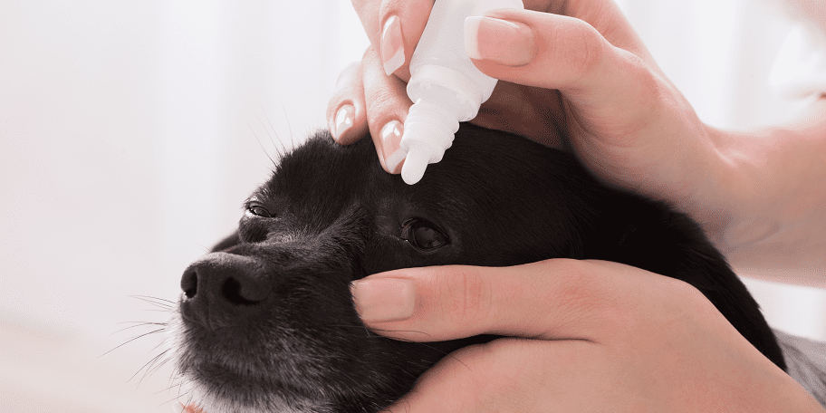
Yellow, or green, discharge from the eyes is usually a sign of infection and needs prompt veterinary attention.
Dry eye is a condition where fewer tears are produced. To compensate, the body instead produces mucous. However, excessive mucous production in the form of eye discharge, can make the eyes red and sore. In this case, there is more probability of ulcers and infections developing.
The mucous produced with dry eye is usually greyish white and if left untreated it can result in a great deal of pain for the dog and ultimately, blindness.
Conclusion
While eye conditions are fairly common in dogs, it is important to carefully monitor any unusual appearance in your dog’s eyes especially when this involves eye discharge.Many conditions are easily treatable, but left untreated, some can cause irreversible damage to the eye.
This Holidays4Dogs article is for information purposes only. Always consult your veterinary surgeon about all matters relating to your pet’s health.